

![]()

![]()
![]()
![]()
![]()
![]()
![]()
-Electronic FHR montoring and Non-stress test
This website is part of the comprehensive online
Obstetric and Gynecology Atlas and Gallery
Hundereds of carefully categorized obgyn illustrations, and real life ultrasound scan images and clips from clinical practice with desription, user comments lightbox..etc.

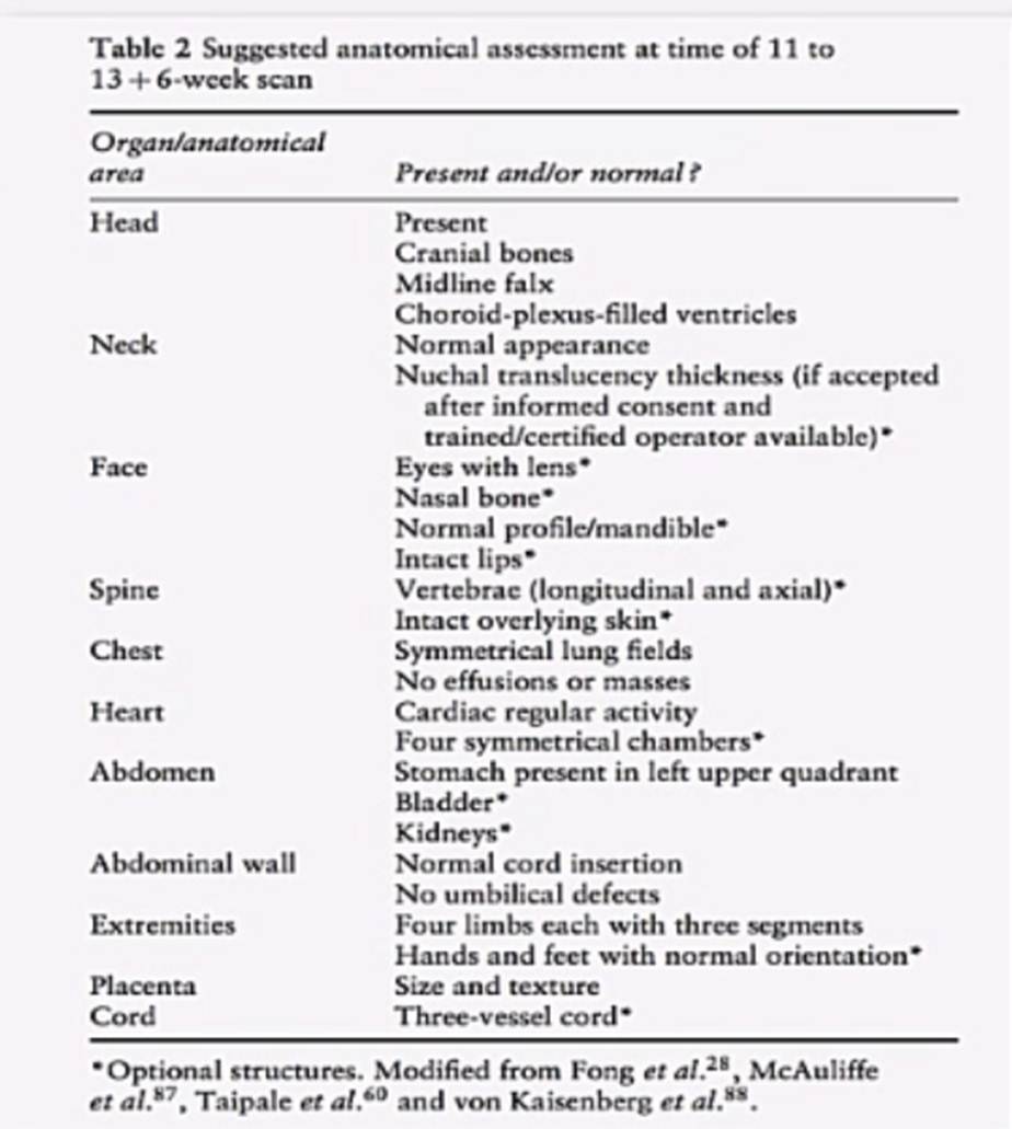
11 weeks to 14 weeks scan
Tip: Examination MUST be done with full urinary bladder.


Illustration of Nuchal thickness mesurement, much more images and clips of 11 to 14 weeks anomaly scan are available in OBGYN Atas and Gallery
Nasal bone Fetal nasal bone is seen in the saggital view at 11 to 14 weeks gestation as it is easier to view at this gestational age. Absent nasal bone (flat face) may reflect chromosomal anomaly. I find this when associated with increased NT is an indication to perform free fetal DNA testing in maternal plasma.

Illustration of Fetal Nasal bone visulization, much more images and clips of 11 to 14 weeks anomaly scan are available in OBGYN Atas and Gallery

Illustration to compare normal feindings with increased nuchal thickness and absent fetal nasal bone, much more images and clips of 11 to 14 weeks anomaly scan are available in OBGYN Atas and Gallery
BASIC STRUCTURAL SCAN
WILL BE REVIEWED ALONG THE RELEVANT SPECIFIC SECTIONS
CRL; crown-rump length; between 7 and 13 weeks of gestation CRL is very important in order to correctly determine EDD (Expected Delivery Date). It is the most accurate measurement for dating pregnancy and can be used later in the management of FGR (Fetal growth restriction).
It is measured as the largest dimension of embryo/fetus, excluding the yolk sac and extremities.
Gestational age estimation is most accurate by CRL measurement, it carries less than 5 days falacy while 2nd trimester BPD carries about 7 days falacy, third trimester scan is not accurate to estimate gestational age.


Illustration of Crown-rump length and Biparietal diameter measurement, much more images and clips of 11 to 14 weeks anomaly scan are available in OBGYN Atas and Gallery
Cervical Measurement: Only with a full urinary bladder the cervical length and internal os diameter are measured and documented. These measurements are correlated with the patient’s obstetric history.
- Fetal Heart Rate/rhythm
- Fetus growth: appears large or small for the stage of pregnancY
- Decreased fetal movements during scan
- Absent fetal abnormalities
Copyrights © Dr.Amr Essam 2018 - 2020